Current topics
Two distinct actin waves correlated with turns-and-runs of crawling microglia
Freely crawling cells are often viewed as randomly moving Brownian particles but they gener- ally exhibit some directional persistence. This property is often related to their zigzag motile behaviors that can be described as a noisy but temporally structured sequence of “runs” and “turns.” However, its underlying biophysical mechanism is largely unexplored. Here, we care- fully investigate the collective actin wave dynamics associated with the zigzag-crawling movements of microglia (as primary brain immune cells) employing a number of different quantitative imaging modalities including synthetic aperture microscopy and optical diffraction tomography, as well as conventional fluorescence imaging and scanning electron micros- copy. Interestingly, we find that microglia exhibit two distinct types of actin waves working at two quite different time scales and locations, and they seem to serve different purposes. One type of actin waves is fast “peripheral ruffles” arising spontaneously with an oscillating period of about 6 seconds at some portion of the leading edge of crawling microglia, where the vigor- ously biased peripheral ruffles seem to set the direction of a new turn (in 2-D free space). When the cell turning events are inhibited with a physical confinement (in 1-D track), the peripheral ruffles still exist at the leading edge with no bias but showing phase coherence in the cell crawling direction. The other type is “dorsal actin waves” which also exhibits an oscil- latory behavior but with a much longer period of around 2 minutes compared to the fast “peripheral ruffles”. Dorsal actin waves (whether the cell turning events are inhibited or not) initiate in the lamellipodium just behind the leading edge, travelling down toward the core region of the cell and disappear. Such dorsal wave propagations seem to be correlated with migration of the cell. Thus, we may view the dorsal actin waves are connected with the “run” stage of cell body, whereas the fast ruffles at the leading front are involved in the “turn” stage. PLoS ONE (2019) 14(8): e0220810.

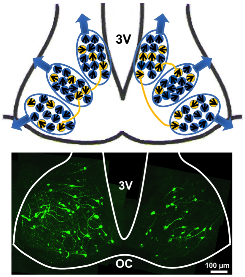
Diversity in the structure of action potential‐mediated neural connectivity within rat supra chiasmatic nucleus
Action potential (AP)‐mediated cell‐to‐cell communication is essential for the fre- quency‐locking and phase‐synchronization of the clock cells within the biological master clock, suprachiasmatic nucleus (SCN). Nevertheless, the morphology of its network connectivity is largely unexplored. Here, with an optimized optogenetic light‐stimulation and scanning protocol, we report some key characteristics of the in- hibitory receptive field (IRF), the area which brings inhibitory synaptic currents to a given target cell, and basic statistics of the inhibitory network connections of rat SCN clock cells. ChR2 transfected, slice cultures of rat SCN were stimulated by a blue power LED light in a repetitive box‐scanning modes, while a target cell was whole‐ cell patched. The registered inhibitory postsynaptic currents, which were brought by light‐induced APs of presynaptic neurons, were mostly GABAergic. The sizes and shapes of IRFs of SCN cells were very diverse, and the number of presynaptic cells making up the IRF of a given target cell followed an exponential distribution with an average value of 8.9 approximately, according to our clustering analysis which is based on a hybrid measure D, combining the physical distance r and the difference in the current amplitudes of two different sites. Although this estimate inevitably depends on the construct of the measure D, it is found not so sensitive on the pa- rameter w, which weighs the relative significance of the current amplitude different with respect to the physical distance r: The average number of presynaptic neurons varies < 26% over a significant range of 0 < w < 30. On average, the presynaptic connection number density around a target cell falls off as an exponentially decreas- ing function of r. But, its space constant (~210.7 μm) is quite large that long‐range (>210.7 μm) neural connections are abundant (>66.9%) within the SCN. Eur J Neurosci (2019);1–16.

Encoding information into autonomously bursting neural network with pairs of time-delayed pulses
Biological neural networks with many plastic synaptic connections can store external input information in the map of synaptic weights as a form of
unsupervised learning. However, the same neural network often produces dramatic reverberating events in which many neurons fire almost
simultaneously – a phenomenon coined as ‘population burst.’ The autonomous bursting activity is a consequence of the delicate balance between
recurrent excitation and self-inhibition; as such, any periodic sequences of burst-generating stimuli delivered even at a low frequency (~1 Hz) can easily suppress the entire network connectivity. Here we demonstrate that ‘∆t paired-pulse stimulation’, can be a novel way for encoding spatially-distributed
high-frequency (~10 Hz) information into such a system without causing a complete suppression. The encoded memory can be probed simply by
delivering multiple probing pulses and then estimating the precision of the arrival times of the subsequent evoked recurrent bursts.
SCIENTIFIC REPORTS (2019) 9:1394


Evaporation-driven convective flows in suspensions of nonmotile bacteria
We report a novel form of convection in suspensions of the bioluminescent marine bac- terium Photobacterium phosphoreum. Suspensions of these bacteria placed in a chamber open to the air create persistent luminescent plumes most easily visible when observed in the dark. These flows are strikingly similar to the classical bioconvection pattern of aerotactic swimming bacteria, which create an unstable stratification by swimming upwards to an air-water interface, but they are a puzzle since the strain of P. phosphoreum used does not express flagella and therefore cannot swim. When microspheres were used instead of bacteria, similar flow patterns were observed, suggesting that the convective motion was not driven by bacteria but instead by the accumulation of salt at the air- water interface due to evaporation of the culture medium. Even at room temperature and humidity, and physiologically relevant salt concentrations, the water evaporation was found to be sufficient to drive convection patterns. To prove this hypothesis, experiments were complemented with a mathematical model that aimed to understand the mechanism of plume formation and the role of salt in triggering the instability. The simplified system of evaporating salty water was first studied using linear stability analysis, and then with finite element simulations. A comparison between these three approaches is presented. While evaporation-driven convection has not been discussed extensively in the context of biological systems, these results suggest that the phenomenon may be broadly relevant, particularly in those systems involving microorganisms of limited motility. PHYSICAL REVIEW FLUIDS 3, 123102 (2018)

Senescent tumor cells building three-dimensional tumor clusters
Cellular senescence, a permanent cell-cycle arrest, is a common yet intriguing phenomenon, in which its beneficial significance for biological
organisms has only begun to be explored. Among others, senescent cells are able to transform tissue structures around them. Tumor cells, whose hallmark is their ability to proliferate indefinitely, are not free from the phenomenon. Here, we report a remarkable observation where senescent cells in a
dense mono-layer of breast cancer colony act as aggregating centers for non-senescent cells in their vicinity. Consequently, the senescent cells actively form localized 3D cell-clusters in a confluent 2D tumor layer. The biophysical mechanism underpinning the surprising phenomenon primarily involves
mitotic cell-rounding, dynamic and differential cell attachments, and cellular chemotaxis. By incorporating these few biophysical factors, we were able
to recapitulate the experimental observation via a cellular Potts Model.

How is the biological master clock organized and functionally connected to generate a coherent 24-hour circadian rhythm?
The suprachiasmatic nucleus (SCN) of the hypothalamus is the biological master clock that governs the daily rhythms in mammals. The individual clock cells of the SCN are quite heterogeneous, in particular, with respect to their intrinsic periods as well as phases. Therefore, they must form a cohesive network through some intercellular coupling to produce a coherent “frequency-locked” 24hr rhythm. Interestingly, these very small (< .5 mm diameter) SCN nuclei support self-organized phase waves, similar to the 'cardiac waves' of the heart. We are investigating various cell-to-cell coupling mechanisms and functional connectomes of the SCN clock cells based on different imaging modalities as well as opto-genetic stimulation tools. We have recently demonstrated that there exist extensive sub-networks of clock cells that share the same circadian phase. Scientific Reports (2016) 6:21463

Learning and memory of in vitro neuronal system accessed by the dynamics of synchronized bursts
Understanding distributed nonlinear dynamical activity of neuronal networks is essential in deciphering how the brain performs cognitive tasks and represents, stores, and processes information. Weare accessing how neural learning and memory operate, in particular, from the viewpoint of synchronized neural bursts. Neuronal networks in culture exhibit synchronized activity of bursts of spikes, and they seem to be a generic property of any growing network of neurons in development. We have recentlydemonstrated that the in vitro model system can alter its bursting dynamics following patterned stimulations and itis reverserable. In addition, we have shown that there exist deterministic features in seemingly random sequence of spontaneously-generated synchronized bursts.
Kim June Hoan, Heo Ryoun, Lee Ho Joon
Density waves in proliferating tumor tissue
Our recent investigation shows a large-scale periodic wave activity in expanding tumor tissue, which is visually similar to a dense population of chemotaxing dicty
amoeba. Shown below is an exemplary collective ratcheting movements of tumor cells forming a large circular wave moving towards the center (marked by “x”), overlaid on the top of a phase-contrast image. New J. Phys. 18 (2016) 103032
Yang Taeseok D., Kim Hyun

How haptotactic cell-to-cell interaction can enhance chemotactic cell aggregation?

We study an interesting aggregation dynamics of mathematical model cells, when they perform chemotaxis in response to an externally imposed global chemical gradient while they influence each other through a haptotaxis-mediated social interaction, which confers intriguing trail patterns. We find that the cell-to-cell interaction confers a far more compact aggregation resulting in a much higher peak equilibrium cell density. The mathematical model system is applicable to many biological systems such as swarming microglia and neutrophils or accumulating ants towards a localized food source.
Tae-goo Kwon and Yang Taeseok D.
Cardiac reentries (spiral waves) and their instabilities
Ventricular fibrillation (VF) is one of the most deadly cardiac arrhythmia during which different parts of the heart beat asynchronously, thus the heart cannot pump blood properly. VF is a 'dynamics disease' that is caused by cardiac spiral wave instabilities. During the last several years, our laboratory has pioneered in studying cardiac spiral waves and their transition to 'alternans,' a harbinger of VF, by developing an in vitro cardiac system and bringing in a new phase contrast imaging technique. Continuing on the previous effort, we are exploring cardiac wave instabilities leading to VF and characterize important properties such as 'restitution curve' and 'CV dispersion relation' under various pharmacological conditions.
Who: Heo Ryoun

Single cell motility & collective patterns in populations of biological cells
Freely crawling biological cells, like "dicty" amoeba, often exhibit directional persistence even in the absence of any external cue and itsmotile behavior is orchestrated by spatiotemporal actin dynamics within cell body. On the other hand, these cells often communicate to each other to exhibit some intriguing behaviors. One good example is the population of microglia (the immune cells of the brain) that form complex networks of trails for "self-trafficking." Another good example is the highly coordinated density waves in proliferating tumor cells. We are currently investigating the origins of these patterns in model simulations as well as in experiments.
Who: Yang T (Daniel), Kwon Tae Goo, Park Jin-sung, Kim Hyun

Calcium waves in network of astroglia and their interactions
In neuron-glia co-culture systems, populations of astro-glia often exhibit 'calcium transients' that match with the neural bursts almost in one-to-one fashion. The glia can support interesting calcium wave activities as well. On the other hand, a growing set of evidences augments the role of glia as regulatory agents of neuronal activity, undertaking active roles in brain neural computation. Therefore, we are interested in characterizing modeling spontaneous and induced (electrical and calcium) spatiotemporal activities that arise in co-cultures of neurons and glia, and associate them with the learning ability of the neural circuits. We have recently found thatalocalized extrinsic ATP perturbation can induce a back-propagating calcium wave in high-density cultures of astrocytes.

Who: Min C. H.